Papillary Thyroid Microcarcinoma– specific ultrasound findings showing factors leading to high risk of progression.
Papillary Thyroid Microcarcinoma– specific ultrasound findings showing factors leading to high risk of progression.
- Seoul National University Hospital announces analysis results of 699 patients with Papillary Thyroid Microcarcinoma under active surveillance
- If ultrasound findings show diffuse thyroid disease + increased blood flow within the tumor, the risk of tumor progression increases by 3.5 times.

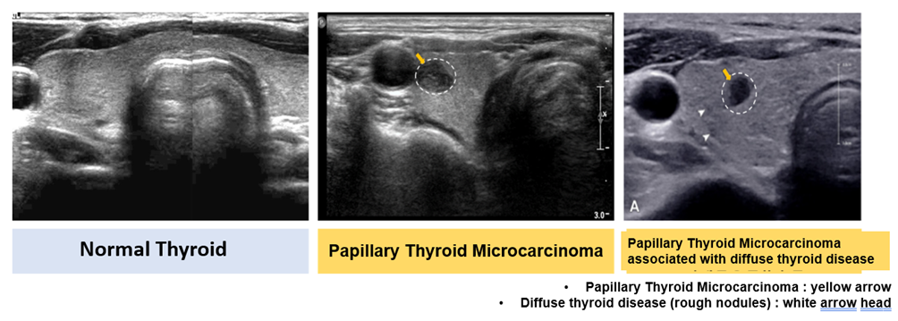
[Photo] Comparison of thyroid ultrasound (from left: normal thyroid, papillary thyroid microcarcinoma, papillary thyroid microcarcinoma with diffuse thyroid disease)
‘Papillary Thyroid Microcarcinoma’, where the tumor is smaller than 1cm, has a good prognosis, so only active follow-up observation is performed instead of surgery. However, domestic researchers have recently confirmed that certain findings on ultrasound may increase the risk of cancer progression.
The research team of Professors Kim Ji-hoon and Lee Ji Ye of the Department of Radiology and Professor Park Young Joo of the Department of Endocrinology and Metabolism at SNUH investigated patients with papillary thyroid microcarcinoma registered in the multicenter prospective cohort (MAeSTro) at Seoul National University Hospital, Seoul National University Bundang Hospital, and the National Cancer Center. The results of analyzing the correlation between ultrasound findings and the risk of tumor progression in these cohorts were announced on November 22nd.
Thyroid cancer is a common carcinoma that ranked first in domestic cancer incidence in 2020. 80-90% belong to papillary thyroid cancer with a high degree of cancer cell differentiation, and among them, ‘papillary thyroid microcarcinoma’, where the tumor is smaller than 1 cm, progresses slowly and has a very low mortality rate. Therefore, domestic and international thyroid societies have stipulated that active observation through ultrasound examination can be considered instead of surgery, and in fact, the number of patients opting for this is increasing.
To evaluate whether active surveillance is appropriate for a patient, it is important to predict the long-term prognosis and progression rate of the tumor, but the risk factors for papillary microthyroid carcinoma have not been clearly identified so far.
The research team followed 699 patients with papillary thyroid microcarcinoma who underwent ultrasound examinations more than twice as part of active surveillance for a median of 41 months and analyzed the correlation between ultrasound findings and tumor progression. Tumour progression was assessed by tumor size increase, extrathyroid tissue invasion, and lymph node metastasis.
As a result, two ultrasound findings, ‘diffuse thyroid disease’ and ‘increased intratumoral blood flow’, were found to be independently associated with tumor progression. Diffuse thyroid disease refers to a condition in which the thyroid parenchyma appears uneven on ultrasound or the blood flow is generally increased.
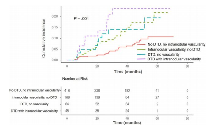
After 4 years of follow-up, the tumor progression rate in patients with two ultrasound findings simultaneously was 21% (10 out of 48 patients). On the other hand, the tumor progression rate in patients without these findings was only 6% (25 out of 418).

[Graph] Kaplan-Meier curve shows time-dependent cumulative incidence of tumor progression in participants with papillary thyroid microcarcinoma. The group with only one finding had a 2.2 times higher risk of tumor progression than the group with no findings. The group that showed both findings simultaneously had a 3.5-fold higher risk of tumor progression. In other words, subgroups classified based on ultrasound findings accurately stratified the risk of tumor progression.
As a result of the risk analysis, compared to patients without diffuse thyroid disease and no findings of increased intratumoral blood flow, patients with only one finding had a 2.2 times higher risk of tumor progression. On the other hand, patients who showed two findings simultaneously had a 3.5-fold higher risk of tumor progression.
In particular, if there was a finding of ‘diffuse thyroid disease’, the risk of tumor size increase was 2.7 times higher than in patients without the finding, and if there was a finding of ‘increased intratumoral blood flow’, the risk of lymph node metastasis was about 5 times higher.

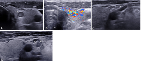
[Picture] A case of papillary thyroid microcarcinoma with increased blood flow and lymph node metastasis. In the colour Doppler image (B), increased blood vessels are observed, and the lymph nodes (C arrows) are small and benign. Results of 12-month follow-up (D) Lymph node metastasis of papillary thyroid microcarcinoma occurred.

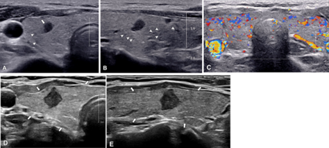
[Picture] Papillary thyroid microcarcinoma accompanied by diffuse thyroid disease, increasing in size. The parenchyma is uneven and rough (A,B arrowheads), and increased blood vessels are observed inside the nodule in the color Doppler image (C). As a result of 18-month follow-up (D, E), the size of microscopic papillary thyroid cancer, which had a maximum diameter of 4 mm, expanded to 8 mm.
The research team emphasized that by considering ultrasound findings related to the progression of papillary thyroid microcarcinoma, the suitability of active observation and the accuracy of assessing the likelihood of progression can be improved.
In addition, clinical characteristics such as young age (less than 30 years old), male sex, and increased levels of thyroid-stimulating hormone (TSH) were also found to be associated with the rapid progression of papillary thyroid microcarcinoma.
Professor Kim Ji-hoon of the Department of Radiology said, “When conducting active surveillance for Papillary Thyroid Microcarcinoma, customized tumor progression surveillance will be possible if the patient's clinical characteristics and ultrasound findings are also evaluated.”
“In addition, verification of results through long-term follow-up data is necessary.”, he added.
The results of this study were published in ‘Radiology’ (Journal of the North American Society of Radiology, IF: 19.7), a prestigious journal in the field of radiology.

[From the left in the photo] Professor Kim Ji-hoon Kim and Lee Ji Ye of the Department of Radiology, and Professor Park Young Joo of the Department of Endocrinology and Metabolism.